St. Louis, Missouri, USA
February, 25 2025

Danforth Center scientists Tessa Burch-Smith, PhD, and Kirk Czymmek, PhD, in collaboration with researchers at the CryoEM Facility at Stanford University, are embarking on a pioneering initiative to develop new cryopreservation and nanoscale imaging procedures for volume electron microscopy (vEM) of plant tissues. This project aims to overcome longstanding challenges in applying these advanced imaging techniques to plant science.
The Danforth Center’s Advanced Bioimaging Laboratory recently acquired a ThermoFisher Scientific Hydra Plasma FIB-SEM, a state-of-the-art technology that leverages vEM for 3D bioimaging of cellular structures. This cutting-edge tool enables researchers to observe plant cells and tissues after cryopreservation, providing unprecedented insights into how cellular components interact and function.
“Volume electron microscopy is revolutionizing our ability to understand the structure and spatial organization of cells and tissues,” said Czymmek, director of the Advanced Bioimaging Laboratory at the Danforth Center. “By combining vEM with cryo-electron tomography (cryoET), we can push the boundaries of in situ structural biology and gain deeper insights into plant cellular processes.”
Advancing Plant Science Through Cryopreservation
Understanding how cells and tissues are organized is fundamental to advancing plant science. While electron microscopy is a powerful tool for studying cells at the nanoscale, proper cryogenic preservation is essential to maintaining sample integrity and avoiding distortions that could impact results.
However, plant cells present unique challenges for cryopreservation due to their physical and chemical properties. As a result, plant science has lagged behind other fields in harnessing structural imaging techniques to explore the inner workings of macromolecular complexes and cellular compartments. This project aims to bridge that gap, unlocking new possibilities for developing crops with enhanced resilience, productivity, and sustainability.
“This work will provide critical insights into plant cell function and help us develop crops better suited to meet global food security challenges,” said Burch-Smith, associate member at the Danforth Center.
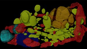
 A 3D reconstruction of the cell contents of the single cell alga Chlamydomonas reinhardtii acquired by cryo-vEM on the Thermo Fisher Scientific Hydra Plasma FIB-SEM in a Department of Energy funded collaborative project between the Czymmek and Zhang Labs at the Danforth Center Advanced Bioimaging Laboratory.
Open-Source Methods and Workforce Development
To maximize scientific impact, the researchers will share their newly developed cryopreservation and imaging techniques in public databases, allowing other scientists worldwide to integrate these methods into their own research.
Additionally, the project includes training early-career scientists in advanced imaging techniques, skills that are in high demand across academia and industry, helping to cultivate the next generation of plant science researchers.
The project is backed by a $1.5 million grant from the National Science Foundation’s Plant Genome Research Program (PGRP), which supports the development of innovative tools, technologies, and resources for plant research.
This initiative builds on the groundbreaking work of Jacques Dubochet, Joachim Frank, and Richard Henderson, who were awarded the 2017 Nobel Prize in Chemistry for their role in developing cryo-electron microscopy. This new research collaboration will further refine three-dimensional visualization of vitrified plant cells, expanding the understanding of plant-microbe interactions in both pathogenic and beneficial contexts.